- Pathogenesis:
- Dogs are the main intermediate hosts of
hydatid disease.
- Eggs get ingested, hatch in the stomach
and duodenum, travel to the liver via portal venous drainage, encyst in
the liver and grow slowly.
- The cysts can exert mass effect on the
surrounding liver and biliary system. The right lobe of the liver is
most commonly involved.
- The cysts can rupture into the pleural
cavity, peritoneal cavity, alimentary canal, or biliary tree, causing
profound shock, peritonitis, and anaphylaxis.
- Radiographic findings:
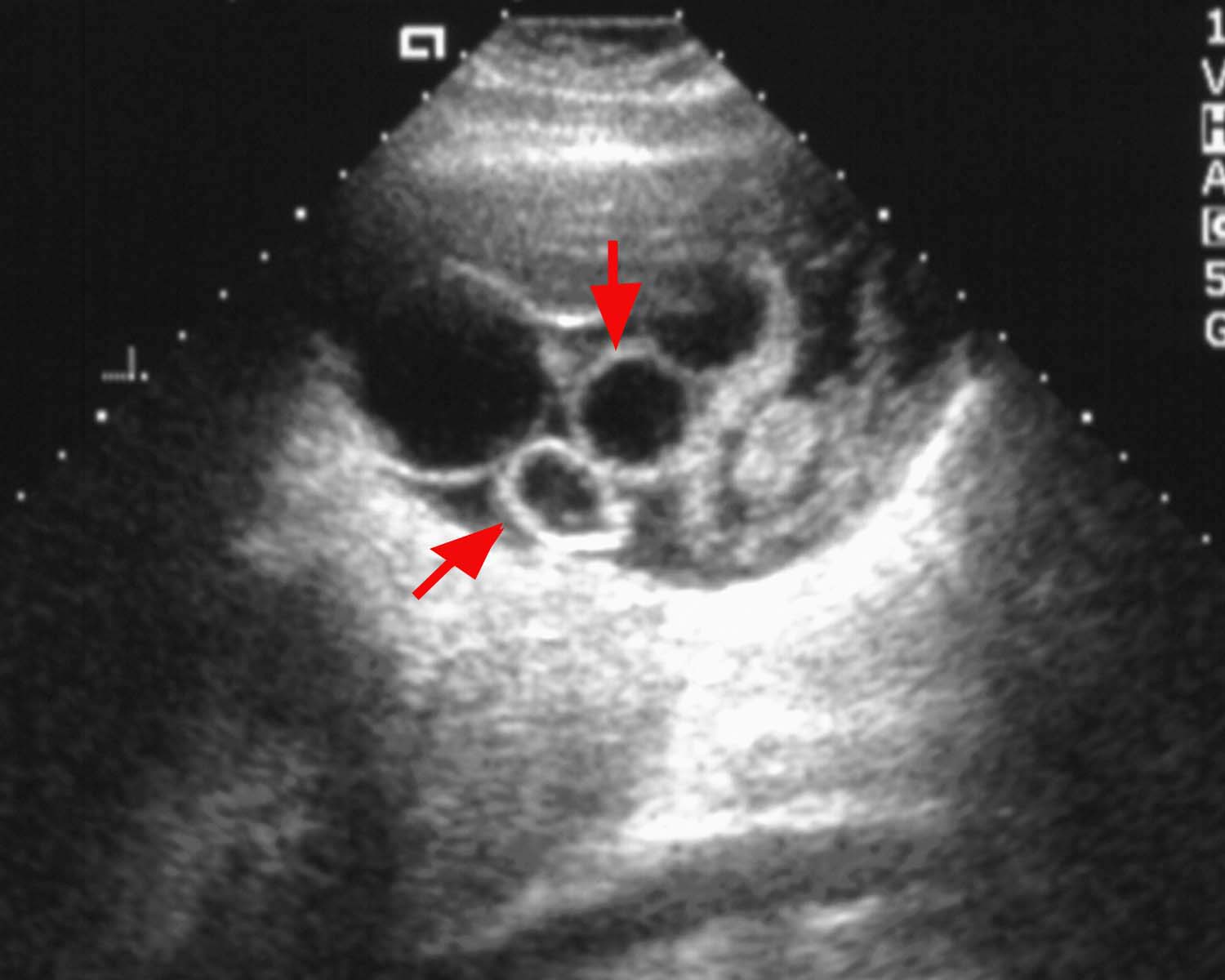
- U/S: double-layered cyst, "classic"
double-line sign, water lily sign, racemose.
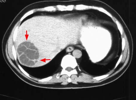
- CT: may see calcified wall, membrane
separation, dependent debris, and/or focal areas of increased attenuation within the cysts (arrows).
|