GI Radiology > Spleen > Infectious > Abscesses
Abscesses: pyogenic vs. fungal
![]()
-
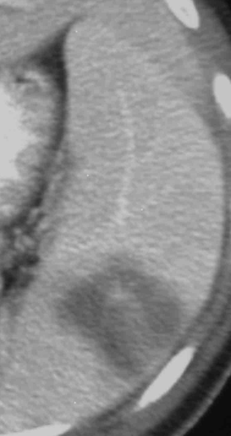
The CT image above shows a rounded hypoenhancing lesion in the spleen, consistent with an abscess. Splenic abscesses usually present in a rounded or lobulated form on CT. Utilizing CT, there is decreased attenuation on unenhanced images and decreased enhancement due to the presence of prurulent material and poor blood flow to the specified area. Subsequent workup after intial detection of a splenic abscess must include a cardiac echocardiogram to rule out bacterial endocarditis. Endocarditis constitutes the primary source of infection due to septic emboli manifest in the spleen.
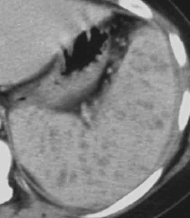
The patient with the above image was a child who presented with ALL. This CT was done with contrast and showed multiple small hypodensities in the spleen. This patient has disseminated Candida infection.
-
This appearance on CT of many small hypodensities could represent an array of pathology, including: metastatic disease, lymphoma, abscesses, multiple hematomas, splenic infarction, traumatic or congenital cysts.
-
In this case, the patient presented with immunosuppression (ALL), so the diagnosis is a systemic fungal disease - candidiasis. Candida is seen in patients with diabetes, AIDS, or hematologic malignancies (like the above patient). It is also seen in patients who are receiving steroid therapy. Lesions from systemic candidiasis are seen in the brain, heart, liver, spleen, and kidneys.
-
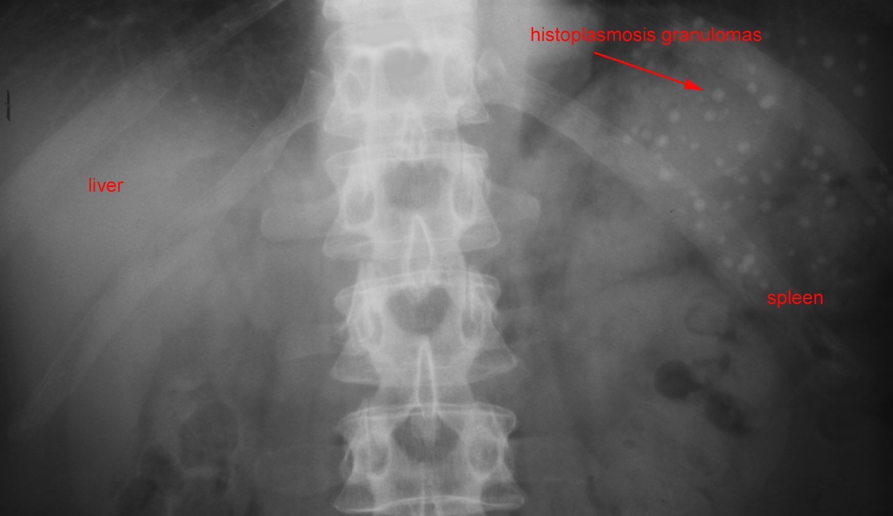
The plain film shown demonstrates numerous round calicifications diffusely distributed specifically within the spleen. The likely etiology of the calcified granulomata is Histoplasmosis, which have this characteristic appearance, in comparison to microabscesses.