Upper G.I. Tract Biphasic-Contrast Exam (cont.)

Method
|
Note that all patient positions
described are in relation to the x-ray table.
|
-
While the patient is
lying supine on the x-ray table, slowly inject 0.2 mg of glucagon intravenously.
(When you are just beginning to learn the procedure and are slow, consider
using 0.3 mg.)
-
Raise the x-ray table to the upright position. The patient stands on the
footrest with his back against the table top. (If patient cannot stand,
elevate head of table 30-45 degrees.) Quickly scan the abdomen with the
fluoroscope to check for contraindications to the study: free air beneath
diaphragm or residual barium from a previous exam.
-
Immediately before using the chilled soda siphon, shake it vigorously and
dispense "bubbly barium" into a 10 oz cup. Only fill the cup 1/2 full. Have the patient hold the cup of
barium in the left hand. (Have the technologist hold the cup if patient is
disabled.)
-
Rotate the patient into a left posterior oblique (LPO) position (to
rotate the spine away from overlapping with the esophagus).
-
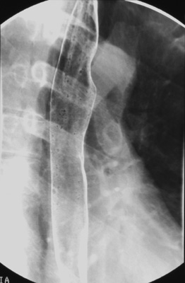
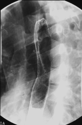
Ask the patient
to drink 1/2 cup of "bubbly barium" quickly and tell him not to belch. Scan
the length of esophagus while the patient swallows. Obtain two
double-contrast spot images - one of the upper and one of
the lower esophagus (including the gastric cardia) during maximal gas distention
after all gas bubbles have disappeared. Collimate the fluoroscopic image side-to-side before taking the esophageal spot images.

-
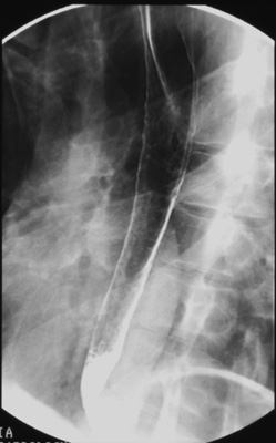
Then, take the empty cup from the
patient and refill it 1/2 full with "bubbly barium". Repeat the same spot images in the
right posterior oblique (RPO) position while the patient holds the cup in
his right hand and drinks an additional 1/2 cup of "bubbly barium". Do not
have the patient stop breathing or swallowing while making the exposures.


|
|
|


![]()
![]()