Upper G.I. Tract Biphasic-Contrast Exam (cont.)

Method
(cont.)
|
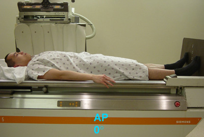
- Take the empty cup from the patient. Turn
the x-ray table into a horizontal position. Give the patient a pillow on
which to rest their head.
-
Ask patient to roll
toward you or rightward (counter-clockwise, as viewed from the foot end
of table) through three 360 degree rotations, stopping in the steep LPO or
left lateral position. (If patient cannot turn around, roll him back and
forth three times from one lateral position to the other.) This is done to
obtain good barium coating of the gastric mucosa while the CO2 is
dissociating from the barium and distending the gastric lumen.

- Take four DC spots in the
following sequence from the distal to the proximal end of the stomach (use
magnification with a 9" or 6" FOV):
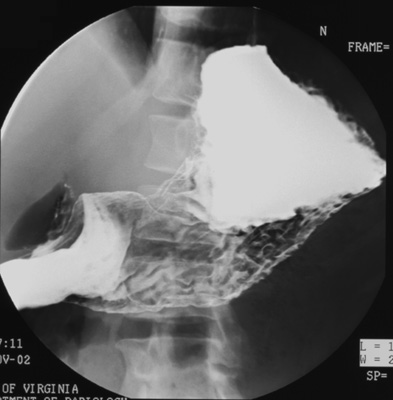
- Gastric antrum (patient LPO)
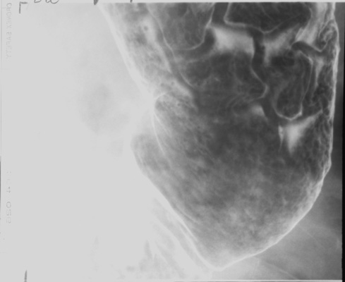
- Gastric body, inferior portion (patient supine, AP)
- Fundus (patient right lateral)
 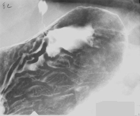
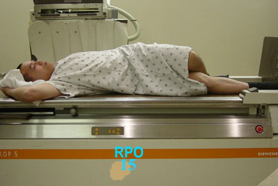
- Gastric body, superior portion (patient RPO). (Elevate
head of table 15 degrees to keep barium from flowing back into gastric fundus as
he rolls back into RPO position.)
- Have patient make another counter-clockwise rotation
(as viewed from foot of table) to refresh the barium coating of the gastric
mucosa. Stop in the steep LPO position.
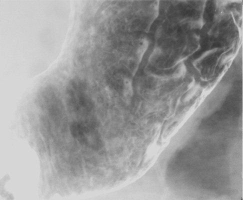
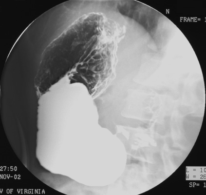
- Take four DC spot images
of the entire stomach using the largest FOV in the following sequence:
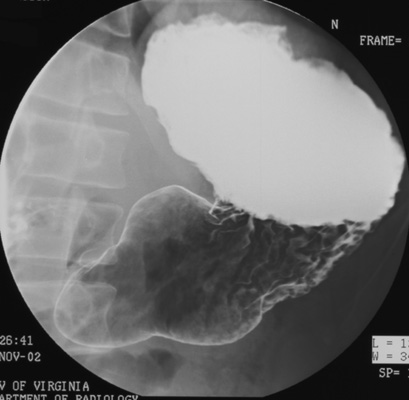
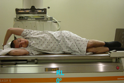
- RPO (First, turn patient into right lateral position
and elevate head of table 15 degrees to keep barium from flowing back into
gastric fundus as he rolls back into RPO position.)
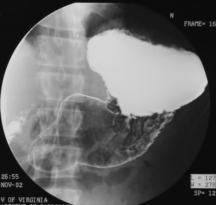
- Right lateral (Wait until duodenal C-loop is
sufficiently filled with barium; otherwise, take this film at end of
study.)
|
|
|

