Upper G.I. Tract Biphasic-Contrast Exam (cont.)

Method
(cont.)
|
-
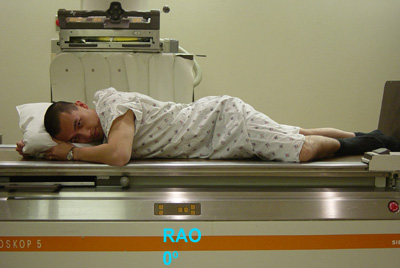
Following the DC examination of the esophagus and stomach, perform
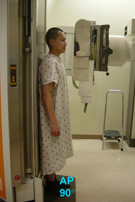
single-contrast graded-compression (SCGC) spots. Turn patient into
right anterior oblique (RAO) position. Place compression paddle beneath
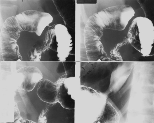
patient and inflate balloon for graded compression of duodenal bulb. Take
one SCGC spot (Zoom to 6" or 9" FOV) of duodenal bulb and one SC spot of
distended proximal duodenal loop.
 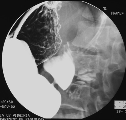
- Turn x-ray table upright. Use compression cone on fluoroscope to
obtain graded compression of duodenal bulb. Take two SCGC spots (Zoom to 6" or 9" FOV) of the bulb.
- Then, use compression cone on fluoroscope and take four SCGC spots (Zoom to 6" or 9" FOV) of:
- Gastric antrum (patient LPO)
- Gastric antrum/body (patient LPO or AP)
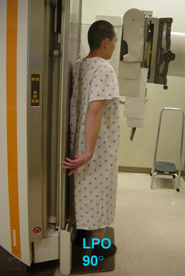 
- Gastric body (patient AP)
- Gastric body/fundus (patient RPO)
- Turn patient into LPO position.
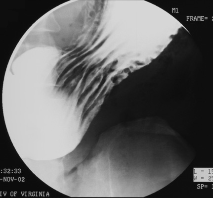
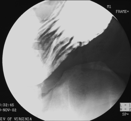
Quickly scan the mediastinum to be sure that the esophagus is empty of
barium. Turn the table into horizontal position and resume fluoro. Take two
DC spots (Zoom to 6" or 9" FOV) of duodenal bulb and two DC spots of
air-filled duodenal C-loop.
- Observe for spontaneous gastroesophageal reflux as you
turn patient towards you (counter-clockwise if viewed from foot of table)
into RAO position.
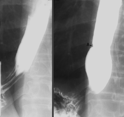
- Return to largest FOV and collimate image from side-to-side. Have the patient drink several
single swallows of dilute, non-carbonated barium through a straw.
Observe esophageal motility and also look for anatomic lesions. Take one
SC spot of the barium-distended lower esophagus and gastric
cardia during breath-holding and one SC spot of the same area during
Valsalva maneuver to evaluate for a possible sliding hiatal hernia.
- Have the technologist take one overhead film (14" x
17", 125 kVp) of the abdomen with patient in prone position.

|
|
|

