Head CT > Degenerative > Pick's Disease > Imaging
Pick's Disease - Imaging
![]()
Radiographically Pick's disease, a variant of Frontotemporal dementia, appears as prominent atrophy of the temporal and/or frontal lobes on CT. Sulcal prominence, widening of the Sylvian fissure with atrophy of the insula, inferior frontal and superior temporal lobes, as well as enlargement of the frontal or temporal horns of the lateral ventricles is most evident on MRI. In addition, MR volumetric analysis may show subtle involvement of the orbitofrontal cortex.
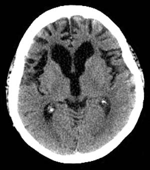 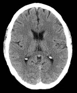 The images above are axial Head CT scans. |
|
On nuclear SPECT cerebral perfusion images, one may see hypoperfusion defects in the ventromedial frontal region in the frontal variant of Frontotemporal dementia. In the temporal lobe variant of Frontotemporal dementia, SPECT demonstrates hypoperfusion in one or both temporal lobes and anterolateral temporal lobe atrophy involving the polar region, fusiform, inferolateral gyri with sparing of the hippocampal formation may be manifested. Invariably the left temporal lobe is more affected than the right temporal lobe. |
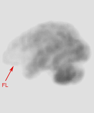 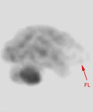 Both images above are SPECT images using Tc-99 in an individual with Pick's disease. |
![]()
![]()