Head CT > Quiz


Quiz

Question 1: Which of the following is NOT true concerning epidural hematoma?

Epidural hematoma is caused by the laceration of a dural artery or venous sinus by a fracture.
The hematoma may cross suture lines.
On CT, the hematoma forms a hyperdense, biconvex mass.
On CT, the hematoma may contain hyperdense foci due to active bleeding.
The hematoma may cross dural reflections.


Question 2: Which of the following is NOT true concerning cerebral contusion?

Cerebral contusion often occurs when the brain impacts an osseous ridge or dural fold.
On CT, cerebral contusions appear as ill-defined hypodense areas.
On CT, cerebral contusions are often mixed with foci of hemorrhage.
After 1 to 2 days, coalescence of petechial hemorrhages into a rounded hematoma is common.
The occipital, temporal, and parietal lobes are the most common locations of cerebral contusion.


Question 3: Which of the following is NOT true concerning diffuse axonal injury?

Diffuse axonal injury can present with a normal head CT.
Diffuse axonal injury is the most common cause of morbidity in CNS trauma.
Diffuse axonal injury is often associated with intraventricular hemorrhage.
On CT, diffuse axonal injury appears as ill-defined areas of high density or hemorrhage.
Acceleration, deceleration, and rotational forces cause diffuse axonal injury.


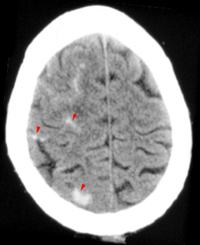
Question 4: Given the following CT, the most likely diagnosis is:


Diffuse axonal injury.
Subarachnoid hemorrhage over cerebral convexities.
Subdural hematoma.
Hypertensive hemorrhage in the occipital lobe.


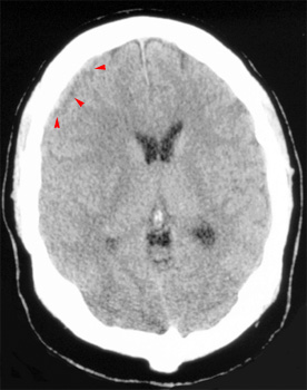
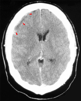
Question 5: The CT on the left, taken prior to contrast administration and the CT on the right, taken after contrast administration, show:


Epidural hematoma.
Subarachnoid hemorrhage.
Glioblastoma multiforme.
Subdural empyema.


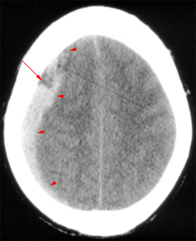
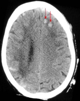
Question 6: Given the following head CT, the most likely diagnosis is:


Acute subdural hematoma.
Epidural hematoma.
Chronic subdural hematoma.
Subarachnoid hemorrhage.


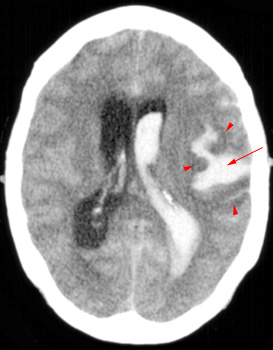
Question 7: Given the following head CT, the most likely diagnosis is:


Hypertensive hemorrhage in the frontal lobe.
Subarachnoid hemorrhage over cerebral convexities.
Diffuse axonal injury.
Intracranial tumors.


Question 8: Which of the following is NOT shown in this CT?


Intraventricular hemorrhage.
Subarachnoid hemorrhage.
Diffuse hypodensity.
Diffuse axonal injury.


Question 9: Which of the following is NOT an advantage to performing a CT scan for stroke?

CT can be rapidly performed.
It is always possible to distinguish between old and new infarcts.
CT allows easy exclusion of hemorrhage.
CT allows the assessment of prenchymal damage.


Question 10: Which of the following is NOT true concerning CT?

CT is the imaging modality of choice for the detecting subarachnoid hemorrhage.
Small subarachnoid bleeds may be inapparent.
On CT, subarachnoid hemorrhage appears as high density within sulci and CSF cisterns.
CT becomes more sensitive days to weeks after the acute phase of a subarachnoid hemorrhage.


Your score is out of 10.

Answers