Pediatric Radiology
> Chest
> Pulmonary Inflammatory Disease
> CXR Findings
Pulmonary Inflammatory Disease

|
Viral Pulmonary Infection - CXR Findings
Bronchitis will manifest on the CXR as peribronchial thickening or "peribronchial cuffing". A bronchus seen on end will show the bronchial wall thickening, and the hilum will demonstrate a dirty appearance, which is well demonstrated on the lateral projection.
The bronchial inflammation results in areas of mucus plugging and atelectasis whereas other areas of the lung will demonstrate hyperinflation from air trapping. The overall lung volumes will be hyperinflated with an increase in the anterior retrosternal space and flattening of the diaphragms.
Viral infections do not have pleural effusions, however, these are relatively common in bacterial infections. The CXR findings for viral infection are the same as that for asthma, which is termed reactive airways disease in the preschool population.
|
 |
 |
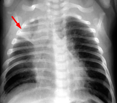
PA CXR demonstrates atelectasis. |
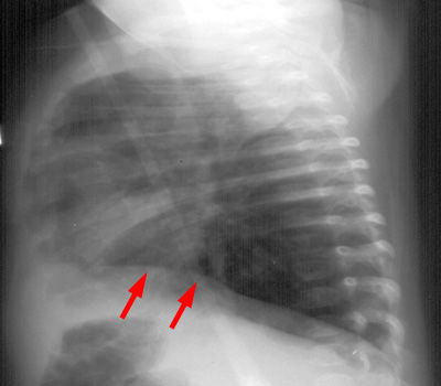
LAT CXR demonstrates flattening of the diaphragms. |
|

