Pediatric Radiology > Chest > Pulmonary Inflammatory Disease > Bacterial Pulmonary Infection
Pulmonary Inflammatory Disease
![]()
Bacterial Pulmonary Infection
Neonatal pneumonia is frequently caused by group b streptococcus and Chlamydia. Chlamydia tends to present slightly later, around 4 weeks, and the chlamydial conjunctivitis is helpful in making the diagnosis. Other common organisms include staphylococcus aureus, haemophilus influenzae type b, and pneumococcus.
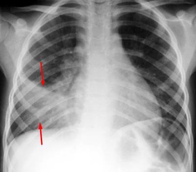
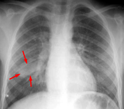
The CXR findings are diagnostic of pneumonia but not specific as to the infecting organism. The most typical presentation is a lobar bronchopneumonia, which manifests on CXR as focal lobar consolidation with air bronchograms. The consolidation may have a round appearance, called "round pneumonia", which can mimic a pulmonary mass.
 |
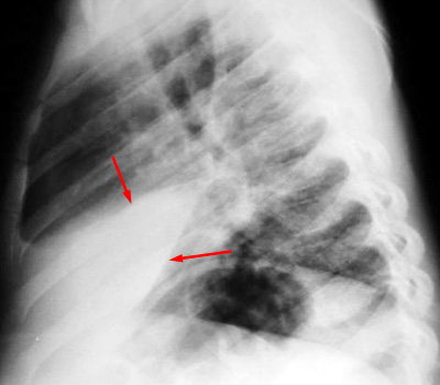 |
| Example of a right middle lobe pneumonia. PA and LAT CXR demonstrate consolidation in the right middle lobe. |
|
 |
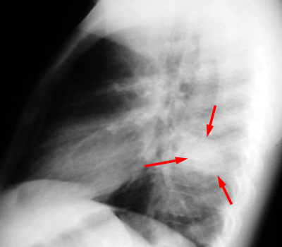 |
| Example of a "round pneumonia." PA and LAT CXR shows a round opacity in the superior segment of the right lower lobe which has the appearance of a mass. |
|
![]()
![]()