Pediatric Radiology > Chest > Pulmonary Inflammatory Disease > Pneumatoceles


Pulmonary Inflammatory Disease

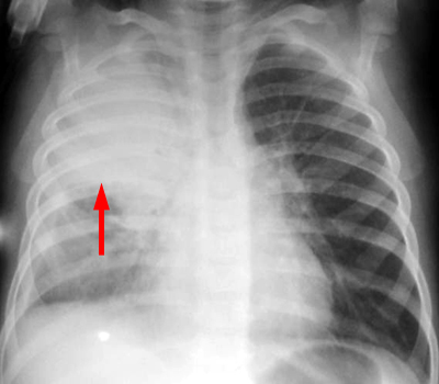
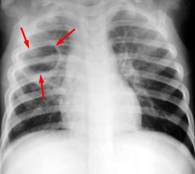
Bacterial Pulmonary Infection


Pneumatoceles are frequent with staphylococcal infections, and they should not be confused with a pulmonary abscess. Pneumatoceles have thin, smooth walls and are seen with an improving clinical picture, whereas pulmonary abscesses have thick, irregular walls with an air fluid level and the child tends to be very ill. Pneumatoceles are thought to be a form of localized pulmonary interstitial emphysema and are self limiting with only the rare case of a large, persisting pneumatocele needing surgery.


Initial CXR shows a dense right upper lobe consolidation.


CXR a week later shows a round cyst with thin walls in the right upper lobe.