|
Clubfoot,
or talipes equinovarus, is a relatively common congenital abnormality
with a frequency of roughly 1:1000. Males are affected twice as often
as females. It is bilateral in about half of the cases. Clinically the foot is plantar flexed, the forefoot is medial and the sole faces inwards. Standard radiographs include AP and lateral weight-bearing films. These XR's are utilized in surgical planning.
The four main components of clubfoot are:
- hindfoot varus
(calcaneus is too far medial)
- equinus heel (fixed
plantar flexion of the heel)
- metatarsus adductus
(adduction of the metatarsals with forefoot varus)
- talonavicular subluxation
Radiographic
characteristics include:
- hindfoot varus - decreased AP talocalcaneal angle, < 20 degrees
- equinus heel - decreased lateral talocalcaneal angle, < 35 degrees (the talus and calcaneus are nearly parallel); increased lateral tibiocalcaneal angle, > 90 degrees
- metatarsus adductus - medial displacement of the first metatarsal relative to the long axis of the talus
- talonavicular subluxation - medial subluxation of the navicular with respect to the talus
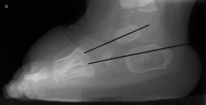
| Talipes Equinovarus
in a 2-year-old male.
A, Frontal view of the right foot shows hindfoot varus with a decreased
AP talocalcaneal angle of 17 degrees. There is also forefoot varus - the line through the long axis of the talus lies lateral to the
first metatarsal and actually bisects the third metatarsal shaft.
B, Lateral view shows a decreased lateral talocalcaneal angle of 11
degrees - the talus and calcaneus are nearly parallel. Equinus
heel is also present with the hindfoot plantarflexed in relation to the tibia. |
|