|
Tarsal
coalition is a congenital fusion of two of the tarsal bones of the foot.
This union can be either a complete or partial bony fusion, or a cartilaginous/fibrous
fusion. Tarsal coalition will often manifest itself as chronic foot pain,
and most patients will present during adolesence.
The two
most common forms of tarsal coalition are:
- calcaneonavicular
(most common) - best seen on oblique view => either a direct connection
(bony coalition) or close proximity with irregular joint margins (fibrous
coalition) can be seen between the calcaneus and the navicular bones;
on lateral view => anterosuperior aspect of calcaneus appears to
extend further than normal ("beaking") towards the navicular
bone.
- talocalcaneal
- more subtle on plain films - CT is often necessary to make the diagnosis;
plain film (lateral) findings include talar beaking, poor visualization
of talocalcaneal joint, and a C-shaped band of overlapping bone noted
over the calcaneus; coronal CT views will show evidence of bony or fibrous
fusion between the middle facet of the talus and the sustentaculum tali
of the calcaneus.
Treatment may include
surgical repair/division of the coalition.
 |
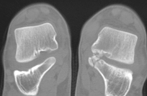
| Talocalcaneal
Coalition in an 11-year-old male. Coronal CT demonstrates
an ossified coalition at the sustentacular articulation of the
calcaneus with the talus. |
|
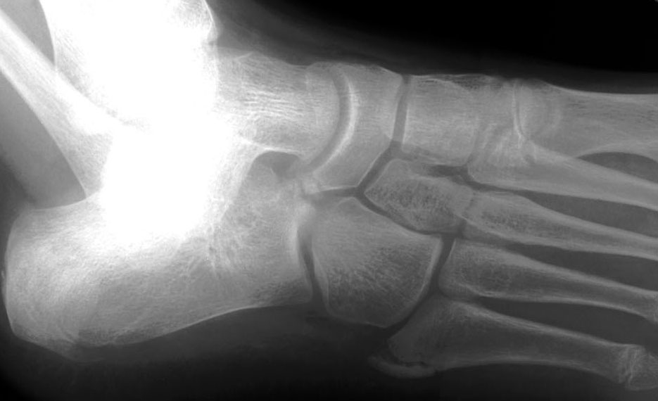 |
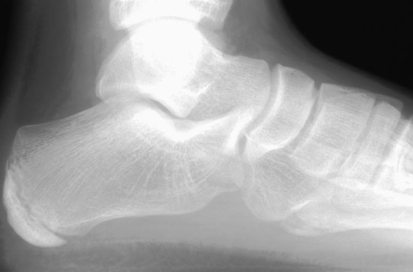
| Calcaneonavicular
coalition in an 11-year old male. Top, Lateral radiograph shows
the superior calcaneus to be elongated. Bottom, Oblique radiograph
demonstrates fibrous coalition with close proximity and irregularity
of the margins of the calcaneonavicular joint. |
|