Pediatric Radiology > Musculoskeletal > Benign Lesions > Distal Femoral Metaphyseal Irregularity
Distal Femoral Metaphyseal Irregularity
![]()
|
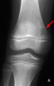
Distal femoral metaphyseal irregularity (cortical desmoid) is an irregular cortical margin with an associated lucency found in the posteromedial aspect of the distal femoral metaphysis. It is thought to be an avulsion off the medial supracondylar ridge of the distal femur. These lesions may or may not be associated with pain. They are primarily found in young boys > young girls (found in up to 10% of boys aged 10-15 years). The lesions are often bilateral. Radiographic findings:
Familiarity with the appearance and location of the lesion as well as the patient's age are helpful in differentiating this benign process from an aggressive malignancy. Obtaining plain films of the contralateral knee to look for bilateral lesions can also aid in determining the benign nature of these lesions, as can a CT. Biopsy should not be performed on these lesions. |
|||
 |
 |
 |
|
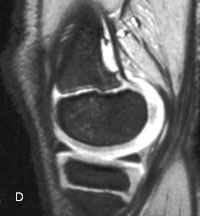
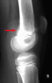
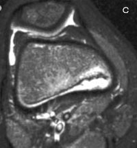
| Distal femoral metaphyseal irregularity in a 7-year-old female. A, Frontal radiogragh shows a well-defined, eccentric lucency (arrow) of the distal medial right femoral metaphysis. B, Lateral radiograph demonstrates a focal cortical irregularity (arrow) in the posterior aspect of the distal femoral metaphysis with adjacent periostitis. C, Axial MRI shows posteromedial cortical irregularity with associated periosteal reaction. D, Sagittal MRI localizes the defect to the femoral origin of the gastrocnemius muscle (medial head). |
![]()
![]()