Pediatric Radiology > Musculoskeletal > Benign Lesions > Benign Cortical Defect
Benign Cortical Defect
![]()
|
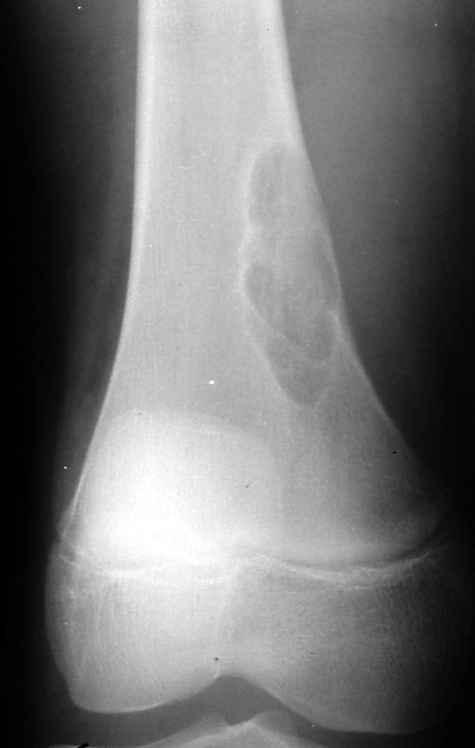
Benign cortical defects are seen in up to 40% of all children at some time in their development. They are most prevalent in children between ages 4-6. The term non-ossifying fibroma is used for lesions >2 cm. They are found emanating from the cortex of long bone metaphyses. The most common location is the distal femur. Radiographically, these lesions appear as lucent, eccentric, well-defined lesions with a thin, sclerotic border. They are typically round or oval in shape. These lesions are not painful, do not exhibit periostitis, and they usually spontaneously regress over time. |
|

|
Benign Cortical Defect in a 7-year-old male. AP view of the right knee shows a radiolucent defect in the distal femoral metaphysis measuring 2cm X 1cm. Note the slight sclerotic margin surrounding the lucency. |
 |
Non-ossifying fibroma in an asymptomatic 8-year old male. AP radiograph of the right knee demonstrates a multilocular, expansile, well-defined lytic lesion in the medial supracondylar ridge of the femur. |