Pediatric Radiology > Musculoskeletal > Benign Lesions > Osteoid Osteoma
Osteoid Osteoma
![]()
|
Osteoid osteoma is a benign osteogenic tumor that usually manifests itself in patients during their second decade of life (boys > girls). The patients usually present with pain, which is worse at night and is relieved by aspirin. It is most commonly found intracortically within the metadiaphyses or diaphyses of the long bones of the lower extremity (femur > tibia). It can also present in the hands and feet. Radiographic findings:
Treatment: surgical excision or CT-guided percutaneous ablation. |
||

|
 |
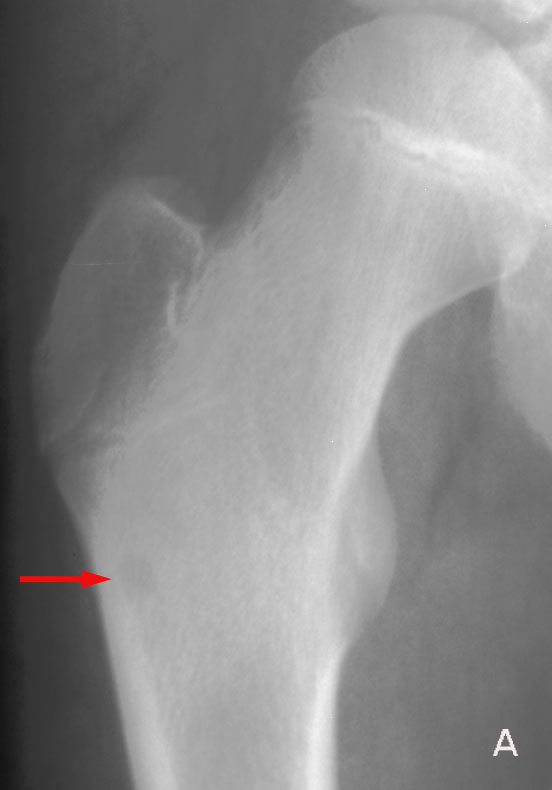
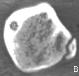
Osteoid Osteoma in a 14-year-old male with hip pain. A, AP radiograph of the right hip reveals a 1cm round, well-defined lytic lesion (arrow) just inferior to the greater trochanter. Note the associated sclerosis in the intertrochanteric region. B, CT demonstrates a calcified central nidus within the lytic lesion. Again, appreciate the sclerotic cortical margin. |