Pediatric Radiology > Musculoskeletal > Benign Lesions > Osteochondroma
Osteochondroma
![]()
|
Osteochondroma is a painless, slow growing osteocartilaginous exostosis (cartilage-capped bony projection). Boys are affected twice as often as females. Growth arises from the metaphyses and the cortex of the lesion is continuous with the adjacent bone. Growth continues until the growth plate closes. These lesions are typically located on the tibia, femur and humerus and often will grow away from the joint. Multiple hereditary exostoses (osteochondromatosis) is an autosomal dominant disease characterized by multiple, usually sessile, osteochondromas. The knee, ankle and shoulder are simultaneously affected. Complications:
|
|
 |
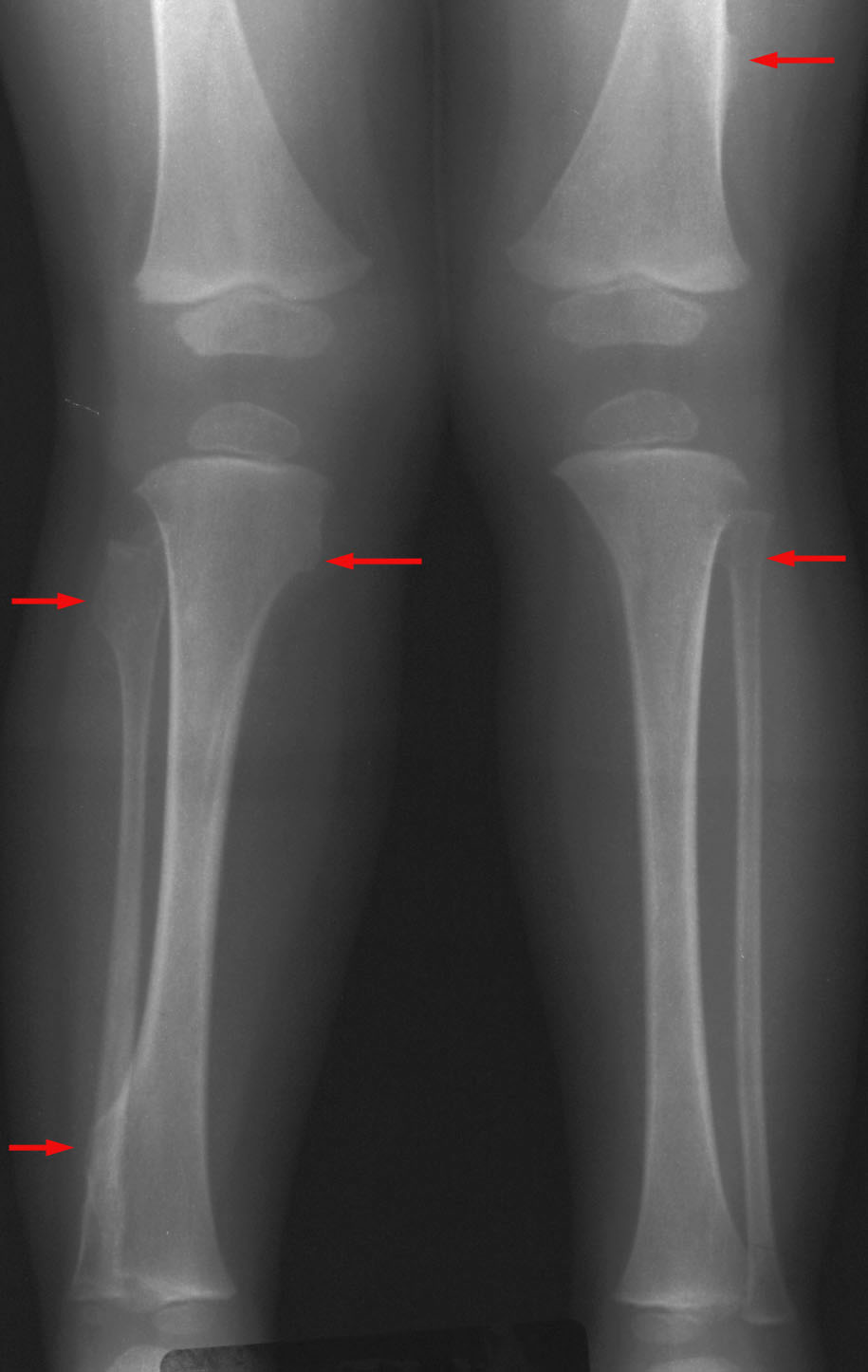
Hereditary multiple exostoses in a 2-year-old female with multiple hard painless "lumps" near multiple joints. AP radiograph of the bilateral lower extremities shows exostoses (arrows) in the distal left femur, proximal left fibula, proximal right tibia and fibula, and distal right tibia. Note how these lesions tend to point away from the nearest joint. |