Pediatric Radiology > Musculoskeletal > Benign Lesions > Enchondroma
Enchondroma
![]()
|
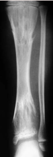
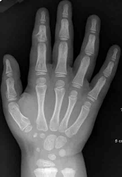
Enchondromas are benign cartilaginous growths of the medullary cavity. Patients are typically 10-30 years old. Enchondromas are the most common benign cystic lesion to involve the phalanges, but they can appear in any bone formed from cartilage. Radiographic findings:
Ollier disease (enchondromatosis) is a non-hereditary condition in which patients have multiple enchondromas. The lesions can be unilateral or bilateral. With growth, these lesions may appear as "flame-shaped" linear configurations that are perpendicular to the physis. Patients often have hand and foot deformities, and are at an increased risk for malignant degeneration to chondrosarcoma. Maffucci disease is a non-hereditary multiple enchondromatosis associated with multiple soft-tissue hemangiomas (phleboliths may be seen in soft-tissue masses on plain film). Patients with this condition have a higher rate of transformation to chondrosarcoma than patients with Ollier disease. Moreover, they are at an increased risk for malignancies of the abdomen and CNS.
|
![]()
![]()