Pediatric Radiology > Musculoskeletal > Benign Lesions > Fibrous Dysplasia
Fibrous Dysplasia
![]()
|
Fibrous dysplasia is a developmental disorder in which medullary bone is replaced by fibrous tissue and abnormal bone. It typically occurs in children between the ages of 5 and 20 and is often associated with endocrine disorders like hyperthyroidism and hyperparathyroidism. This disorder is associated with a very low risk of malignant transformation. There are two types of fibrous dysplasia:
McCune-Albright Syndrome - triad of features include:
|
||
 |
|
|
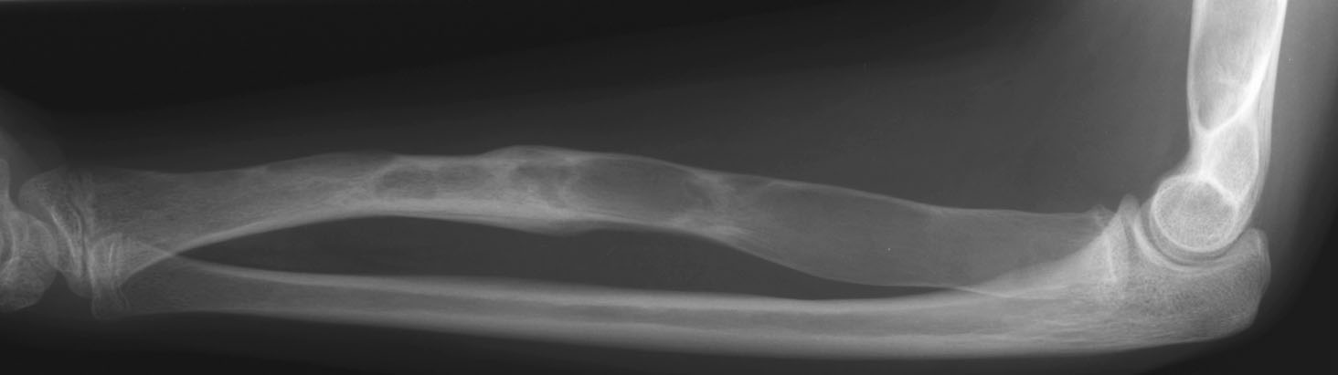 |
| Polyostotic fibrous dysplasia in an 11-year-old female with precocious puberty (McCune-Albright syndrome). Plain film of left forearm shows a "ground glass" geographic lesion in the radius consistent with fibrous dysplasia. Note the healing pathologic fracture in the mid-shaft. A similar ground glass lesion was noted in the patient's left humerus, the distal aspect of which can be seen on this film. |
![]()
![]()