Pediatric Radiology > Musculoskeletal > Aggressive Lesions > Newborn Periosteal Reaction
Newborn Periosteal Reaction
|
Newborn periosteal reaction can result from a host of underlying entities, which are included in the following table:
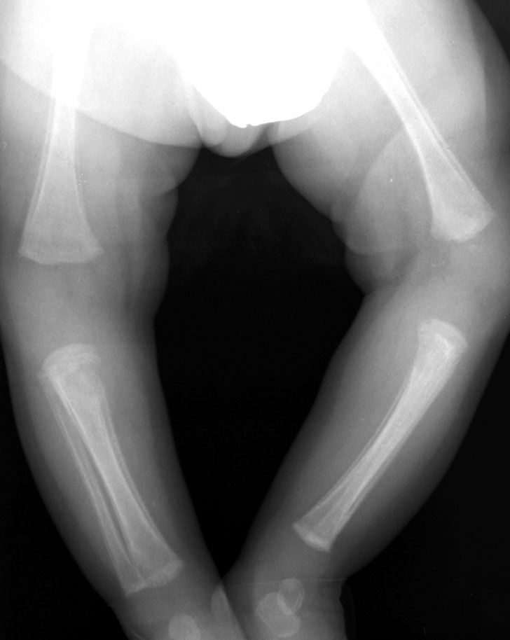
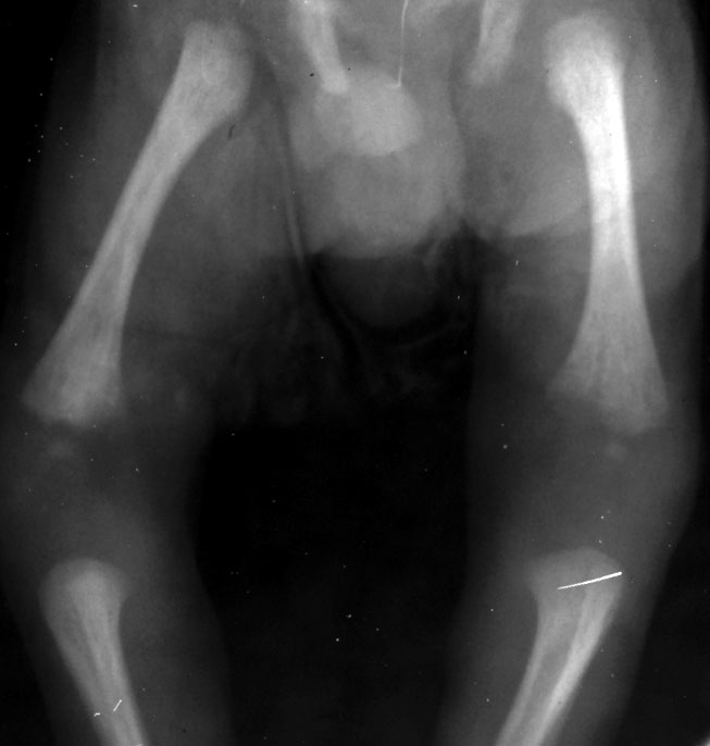
Physiological periosteal new bone formation is seen in up to 1/3 of infants during the first few months of life. This type of periosteal reaction is benign in appearance and usually symmetric, involving the long bones (femur, tibia, and humerus). Radiographs demonstrate one or more dense lines of periosteal reaction along the diaphyses.
Prostaglandins are often used to maintain the patency of the ductus arteriosus in patients with congenital heart disease. Prominent periosteal reaction may be a sequelae from extended use of these therapeutic agents.
The TORCH infections are acquired either transplacentally or at birth. The mnemonic TORCH stands for:
Congenital rubella, acquired transplacentally, has musculoskeletal manifestations in about half of all cases. Radiographic findings include irregular, frayed long bone metaphyses with alternating longitudinal light and dark bands of density (the general appearance is said to resemble a "celery stalk"). Congenital syphilis is also acquired transplacentally (during the 2nd or 3rd trimesters) but musculoskeletal involvement is much more common, roughly 95% of the time. Radiographic evidence of this condition include non-specific metaphyseal lucent bands with periosteal reaction involving multiple long bones. The Wimberger corner sign, thought to be specific to syphilis, is an area of irregular lucency resulting from destruction of the proximal medial tibial metaphysis. Caffey disease (Infantile Cortical Hyperostosis) is an idiopathic condition manifested by periosteal reaction commonly involving the mandible, clavicles, ribs, humerus, ulna, femur, scapula, and radius. This syndrome usually occurs in the first few months of life and is self-limited. Radiographic findings include periosteal new bone formation, sclerosis, and adjacent soft tissue swelling.
|
||||||||
|
|
|
|||||||
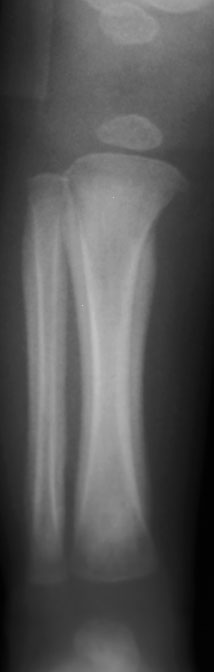 |
Periosteal reaction from prostaglandin therapy in a 1-month old male. AP radiogragh of the distal right lower extremity shows diffuse periosteal reaction involving the tibia and fibula. This patient had a congenital heart disorder that required a patent ductus arteriosus for survival. Prostaglandin E1 was used for patency until surgical correction was performed. |
![]()
![]()