Pediatric Radiology > Musculoskeletal > Constitutional Disorders of Bone > Achondroplasia
Achondroplasia
![]()
|
There are a host of disorders that fall within the category of skeletal dysplasias. These dysplasias are usually categorized by comparing the relative shortening of the proximal versus the distal long bones (e.g. femur versus tibia or humerus versus radius). The various types of skeletal dysplasias include:
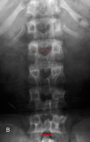
Achondroplasia, the most common of the skeletal dysplasias, is a rhizomelic growth deformity that results from abnormal bone formation at the growth plate. Achondroplasia is an autosomal dominant disorder with a lethal homozygous form and a heterozygous form that demonstrates the multiple clinical manifestations of the disease. Radiographic imaging findings include:
|
|
 |
 |
 |
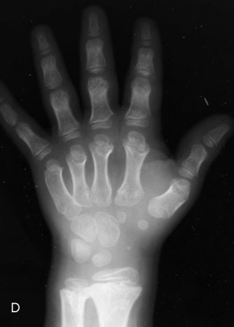 |
| Achondroplasia in a 10-year-old male. A, AP radiograph of the pelvis demonstrates rounded iliac wings with decreased acetabular angles ("tombstone" appearance). B, AP view of the spine shows a decrease in the interpedicular distance from the upper (L1) to the lower (L5) lumbar spine. C, Lateral radiograph of the skull reveals a large calvarium with a diminuitive skull base. D, Radiograph of the right hand shows short, broad tubular bones. |
![]()
![]()