Pediatric Radiology > Musculoskeletal > Metabolic Diseases > Rickets
Rickets
![]()
|
Rickets is a metabolic abnormality in which a deficiency of Vitamin D leads to failure of normal bone mineralization. This disease is manifested earliest at the sites of rapid growth, namely the knees and wrists. Common causes of rickets include: 1) nutritional (dietary insufficiency), 2) malabsorption, 3) renal disease, or 4) increased requirement for vitamin D. Radiographic findings:
Patients may be susceptible to slipped capital femoral epiphyses as well as pathologic fractures. |
|
|
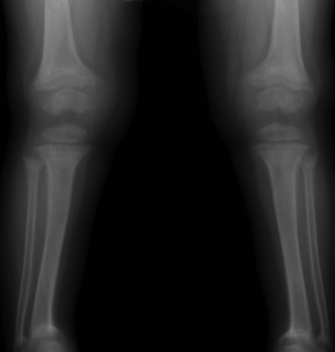
Rickets in a 2-year-old female. AP radiograph of the knees demonstrates widened growth plates and frayed metaphyses. Also note the bowing of the long bones (tibia and fibula bilaterally). |
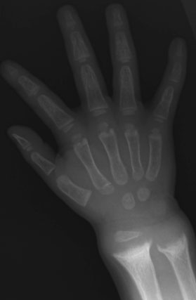 |
Rickets in a 5-year female. Radiograph of the left wrist reveals widened physes. The distal ulnar and radial metaphyses appear cupped and frayed. |
![]()
![]()