Pediatric Radiology > Neurological > Head Ultrasound > Periventricular Leukomalacia
Periventricular Leukomalacia
![]()
|
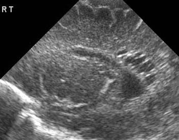
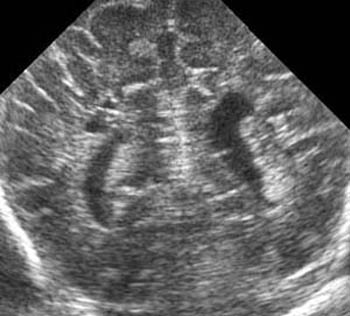
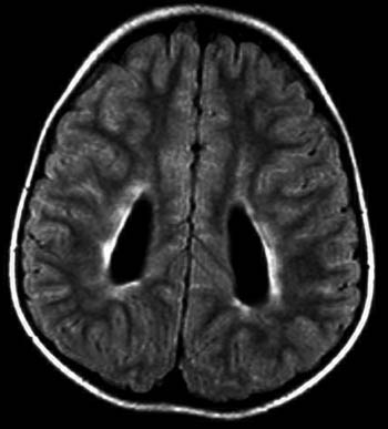
The watershed zone of the premature infant is the periventricular white matter, and perinatal partial asphyxia can result in ischemic damage. Periventricular leukomalacia (PVL) most commonly affects the white matter adjacent to the atria and the frontal horns of the lateral ventricles. PVL is associated with neurologic sequelae such as movement disorders, seizures, and spasticity. Heterogeneous echogenicity is seen within the periventricular white matter and may appear cystic in severe cases. The chronic changes of PVL are best demonstrated with MRI as white matter loss with relative sparing of the overlying cortex. |
|
 |
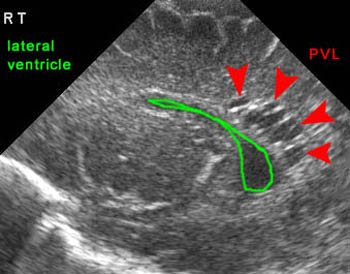 |
| Sagittal head US showing cystic PVL along the right ventricle | |
 |
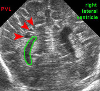 |
| Coronal head US showing cystic PVL along the right ventricle | |
 |
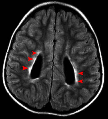 |
| Increased periventricular T2 signal on FLAIR image indicating PVL | |
![]()
![]()