Cardiac MRI > Pathology > Disease of the Aorta and Great Vessels > Aortic Intramural Hematoma
Aortic Intramural Hematoma
![]()
Intramural hematoma (IMH) is a process in which there is rupture of the vaso vasorum into the subintimal space. Intramural hematoma has a high rate of progressing to dissection and is most often the result of long standing hypertension. Unlike an aortic dissection, there is no connection between the aortic lumen and the subintimal space. Like clotted blood elsewhere in the body, the imaging characteristics of an aortic IMH vary according to its age. An acute IMH will appear bright on T2-weighted imaging, but isointense to arterial wall on T1-weighted imaging. Subacute IMH will be bright on both T1- and T2-weighted imaging. Fat saturation techniques can be used to suppress signal from neighboring mediastinal fat.
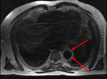
Above is an axial T1-weighted spin echo image. Flowing blood in the descending aorta is dark, but the left side of the aorta has a crescent of bright material (arrows) that represents a subacute intramural hematoma.
![]()
![]()