Cardiac MRI > Pathology > Cardiomyopathies > Arrhythmogenic Right Ventricular Cardiomyopathy
Arrhythmogenic Right Ventricular Cardiomyopathy
![]()
Arrhythmogenic right ventricular cardiomyopathy (ARVC) is a familial cardiomyopathy in which the right ventricular wall is progressively replaced with fibrosis and/or fat. The infiltration with fat/fibrotic tissue can interfere with electrical conductance in the myocardium resulting in ventricular arrhythmias and sudden death. Dilation of the right ventricle and right sided heart failure can occur as well.
Cardiac MRI is the primary imaging modality used to diagnose ARVC. The major imaging criteria for diagnosis of ARVC include severe global or local dysfunction of the RV (with no or only mild LV impairment), global RV dilitation, localized RV or right ventricular outflow tract (RVOT) aneurysm, and fatty infiltration of the RV myocardium (bright on T1, dark with fat saturation sequences). DHE of the right ventricle may suggest infiltration with fibrotic tissue, a finding that would be consistent with ARVC.
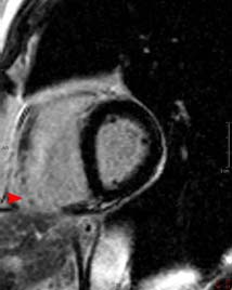
The cine on the top is from a patient with ARVC that shows an aneurysm in the right ventricular outflow tract. The image on the bottom is a DHE image from a different patient with ARVC that shows DHE in the RV wall (arrowhead) that represents fibrosis.
![]()
![]()