Cardiac MRI > Technique > Coronary Angiography Techniques
Coronary Angiography Techniques
![]()
The major impediment to coronary MRA is motion artifacts from breathing and heart motion. While these obstacles have already been discussed, they are more challenging in coronary imaging due to the small size of the vessels. Cardiac motion is at its maximum during mid-systole as the heart contracts and during early diastole as the ventricles fill with blood, thus images are acquired during mid-diastole when heart motion is minimal. Coronary blood flow is also relatively rapid during this time, having the benefit of enlarging the vessels being imaged.
For most cardiac MRI sequences, respiratory motion can be controlled by asking the patient to hold their breath. However, due to the high spatial resolution required, breath hold acquisitions are not possible for most 3-D coronary artery imaging techniques. Navigator imaging techniques can be used to eliminate respiratory motion from the image while still allowing the patient to breathe. With this technique, images are only obtained while the diaphragm is located a specified position across multiple breaths, thus limiting the effects of diaphragm movement.
Many different imaging techniques can be used in coronary MRA including spin echo, gradient echo, SSFP, and gadolinium-enhanced. It is possible to acquire either 2-D or 3-D images.
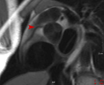
This spin echo image clearly shows the origin of the right coronary artery from the aorta (arrowhead).
![]()
![]()