Emergency Ultrasound > Pelvic Pain > Adenexal Mass
Pelvic Pain - Adenexal Mass
![]()
Clinical
Typical clinical concerns are r/o ovarian cyst/mass/torsion, PID or appendicitis.
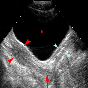
Longitudinal scan through the urine-filled bladder (B) demonstrates a normal adult uterus (red arrowheads) with smooth contours and pear shape. The cervix (red arrow) is recognized at the junction of imaginary lines drawn though the long axis of the uterus and the long axis of the vagina (blue arrowheads).
A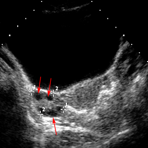 B
B
Normal Ovary. A. Transabdominal. B. Transvaginal. A normal ovary (marked by + cursors) is shown with normal follicles (red arrows) outlining the periphery.
Exam
Start with transabdominal exam with full bladder; then empty bladder and add transvaginal exam if necessary.Obtain multiple sagittal and transverse images of uterus for size and echotexture. Measure the width of the endometrium.If fibroids are present, document their positions. Show and measure ovaries in long axis (sag, AP) and transverse. Measure ovarian cysts that are larger than 2.5 cm. Show a representative long image of both kidneys for size, echogenicity and evaluation of the collecting system.Look for fluid in Morrison's pouch.
Hints for Transvaginal Exam
Having proper image orientation is the key.You should not just rely on the carrot on the screen to tell right from left. If you are in transverse with notch either toward or away from you and if you move your hand toward the patient's left leg, you should simply see more on the left side of the screen (which is the patient's right side). Hit the left/right button if not so.
If you are in sagittal orientation and the notch facing down, and if you push your hand and probe downwards, you should see more on the left side of the screen. Also, by convention, the bladder is always on the upper left of the screen. If not, hit the left/right button.
To stay oriented, think of the transducer as a flashlight. Point the probe towards the area you want to see. Angle up for anterior; angle down for cul-de-sac; angle to the right or left for the adnexae.If you get hopelessly lost, get re-oriented by turning the probe so that the notch on the probe is down (towards the floor) and placing the marker on the left side of the screen.You are now correctly oriented for a sagittal image.Find the bladder.Check the degree of magnification (you don't want too much or too little); check how far the probe is inserted in the vagina (beginners often have the probe in too far or not far enough).
A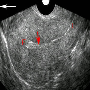 B
B![]()
A. Transvaginal image reveals the uterus fundus (F) and isthmus (I). The red arrow indicates the endometrium and the large white arrow indicates the direction of "up" when scanning transvaginally in longitudinal plane. B. Image of transvaginal transducer.
Sonographic Findings:
1) Functional cyst - smooth, round, anechoic, thin-walled ovarian cyst larger than 2.5 cm.
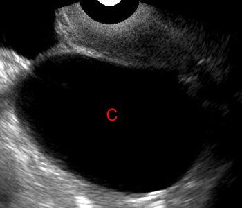
A thin-walled cyst (C) with anechoic internal fluid and size larger than 2.5 cm meets the definition of a functioning ovarian cyst.
2) Hemorrhagic cyst - homogeneous internal echoes, fishnet appearance, retracting clots and fibrous strands, and fluid-fluid levels.
A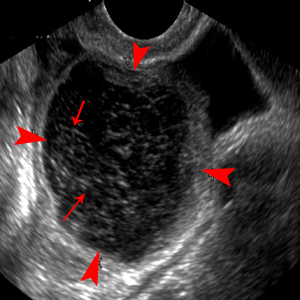 B
B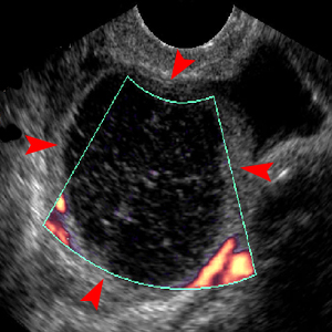
Hemorrhagic Cyst - Fishnet Appearance. A. The cyst (red arrowheads) on the ovary shows fine internal echoes with a fishnet appearance of thin, linear, fibrous strands (red arrows) characteristic of hemorrhage. B. Color Doppler of cyst (red arrowheads) demonstrates lack of internal blood flow characteristic of hemorrhagic cyst.
3) Cystic teratoma - tip of iceberg sign, hyperechoic mass with dark acoustic shadow, and heterogeneous tissues.
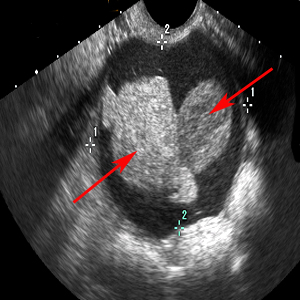
Image of cystic teratoma (between + cursors) with mixed tissues and bizarre solid tissue (red arrows).
4) Endometrioma - adnexal cystic mass with diffuse, low-level internal echoes and hyperechoic foci in the wall.
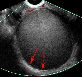
Transverse image of endometrioma contains blood that is higher in echogenicity than most endometriomas. It was initially mistaken for a solid lesion, but color Doppler does not demonstrate internal blood vessels. Echogenic foci in the wall (red arrows) are a subtle but characteristic sign of endometrioma.
5) Ovarian Torsion - diagnosis rests on ovarian enlargement with normal ovarian volume being up to approximately 15 c Other suggestive findings are multiple peripherally based follicles.

Color Doppler image through the ovary (red arrowheads) shows absence of blood flow demonstrating ovarian torsion.
6) Ovarian malignancy - a solid component to an ovarian lesion is the most significant predictor of malignancy; irregular thick wall and septa > 3mm; Doppler demonstration of central blood flow within a solid component.
![]()

Color Doppler of ovary demonstrates blood flow within irregularly thickened septa (red arrows).
![]()
![]()