The following radiographs show the normal anatomy of the elbow.
|
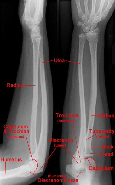 |
Lateral and AP views of normal elbow
|
- A complete exam includes AP, lateral and 2 oblique views.
- The olecranon process of the ulna articulates with the trochlea and olecranon
fossa of the humerus.
- The radial head articulates with the capitulum. These two structures should align
in all projections.
|
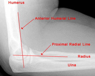 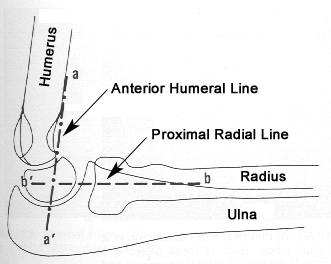 |
Lateral view of elbow with anatomical lines
|
- Anterior humeral line: a line drawn parallel to the anterior humerus should
pass through the middle third of the capitulum.
- Proximal radial line: a line along the longitudinal axis of the radius should
pass through the center of the capitulum in all projections.
|