Emergency Ultrasound > Testicular Pain > Testicular Torsion
Testicular Pain - Testicular Torsion
![]()
Clinical
Testicular torsion must be identified and treated within a few hours to prevent infarction of the testis. Patients prone to torsion lack the normal attachment of the testis and epididymis to the posterior scrotal wall. The patients usually present with sudden onset of severe unilateral scrotal pain. Testicular torsion most often is observed in males younger than 30 years, with most aged 12-18 years. The differential diagnosis also includes epididymitis and orchitis.
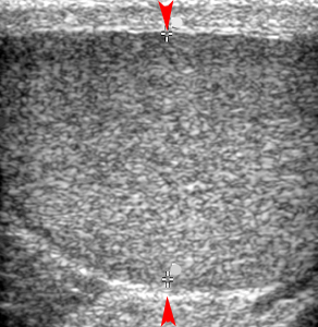
The normal testis (red arrowheads) has a homogeneous, moderately grainy echotexture.
Exam
Begin with the patient in the supine position. Place a white towel below the testicle. Use a second towel to cover the underside of the penis and move it to the lower abdomen so the testicles are easily accessible.
Seeing lack of color flow in a testicle makes the diagnosis of torsion. Always use a 7.5 MHz linear transducer to look at flow in the testicle. Color settings: put on small parts of testicle. The filter must be on 1 or 2 and the color velocity scale on the lowest possible setting for maximum sensitivity. It is absolutely essential to compare to the opposite presumed normal testicle to visualize its vessels so that you know your color settings are right and you are not just getting color noise.
Gray Scale
Obtain long and transverse scans through the scrotum. Include upper, mid, and lower testicle in transverse. Measure the testicles and the epididymis (abnormal is > 1cm). Show hydrocele if present.
Color Doppler
Document color flow to the testicles. Note any increased flow to the epididymis or testicle, which may mean epididymitis or orchitis respectively.
Sonographic Findings:
1)Diffusely hypoechoic torsed testicle compared to the other normal testicle.
A
B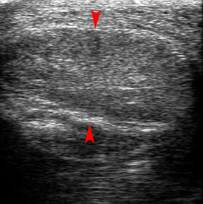
Figure A. The normal testis (red arrowheads) has a homogeneous, moderately grainy, echotexture. Figure B. The torsed testis (red arrowheads) has decreased echogenicity as compared to the normal testis in figure A due to edema.
2) Doppler demonstrates absent or decreased flow in the symptomatic testis compared to the opposite testis.
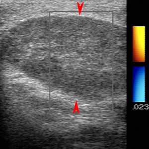
Color Doppler image demonstrates no flow to the painful testis (red arrowheads) characteristic of testicular torsion.
![]()
![]()