Pediatric Radiology > Musculoskeletal > Aggressive Lesions > Osteomyelitis
Osteomyelitis
![]()
|
Osteomyelitis is a bacterial infection of bone that primarily affects infants and young children. 1/2 of all cases occur before 5 years of age with 1/3 of these cases occurring before age 2. Patients may present clinically with pain, fever, and elevated WBC and/or ESR. Many patients will have a history positive for a recent respiratory tract infection or otitis media (most cases of osteomyelitis in the pediatric population have a hematogenous etiology). Osteomyelitis usually occurs in the metaphyses or metaphyseal equivalents in children. Roughly 75% of all infections will involve the long bones (femur > tibia > humerus), while the remaining 25% will involve the flat bones, in particular the hip. Radiographic findings of osteomyelitis:
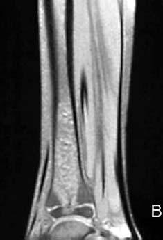
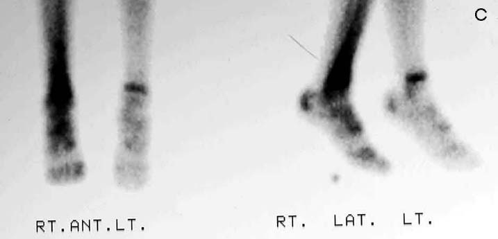
* evidence of osteomyelitis is seen on MRI and bone scans early after the onset of symptoms. |
|
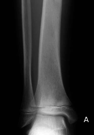 |
Osteomyelitis involving the tibia of a 10-year-old male with foot pain, fever, and elevated ESR. A, AP radiograph of the right leg demonstrates focal demineralization with sclerosis of the distal tibia. B, Sagittal T2WI reveals marrow edema in the distal tibia which crosses the physis to invade the epiphysis. C, Bone scan reveals increased tracer uptake in the right tibia compared with normal uptake on the contralateral side. |
 |
 |
![]()
![]()
© Copyright Rector and Visitors of the University of Virginia 2021