Gas in the gallbladder wall as a result of infection.
A severe condition, usually seen in patients with diabetes.
More common in men.
The sequela of gallbladder ischemia with secondary infection by Clostridia, Escherichia coli, Staphylococcus, or Streptococcus.
Gas present in the gallbladder is formed by above organisms.
These patients have an increased risk of perforation and treatment usually requires emergent cholecystectomy.
Radiographic findings:
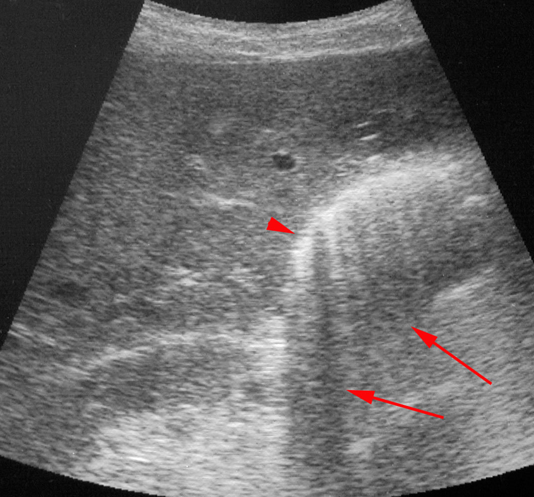
U/S: bright echoes (arrowhead) with poorly defined posterior shadows (arrows) in the wall or lumen.
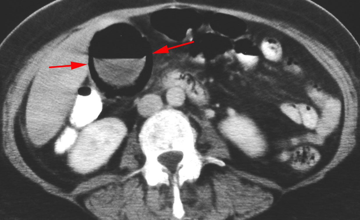
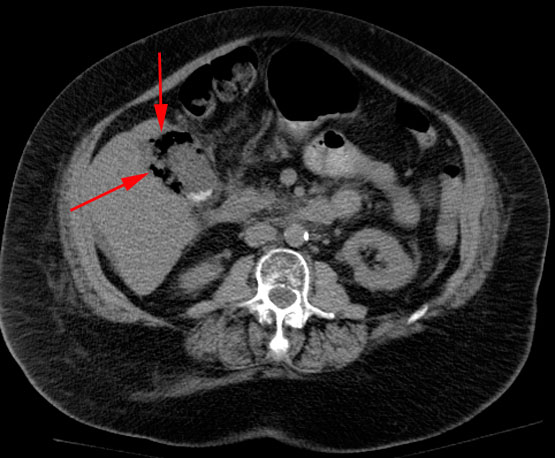
CT: Excellent for detecting gas in the gallbladder wall and lumen, but only performed if plain films are ambiguous. Two separate images below demonstrate gas within the gallbladder lumen and gallbladder wall (arrows).
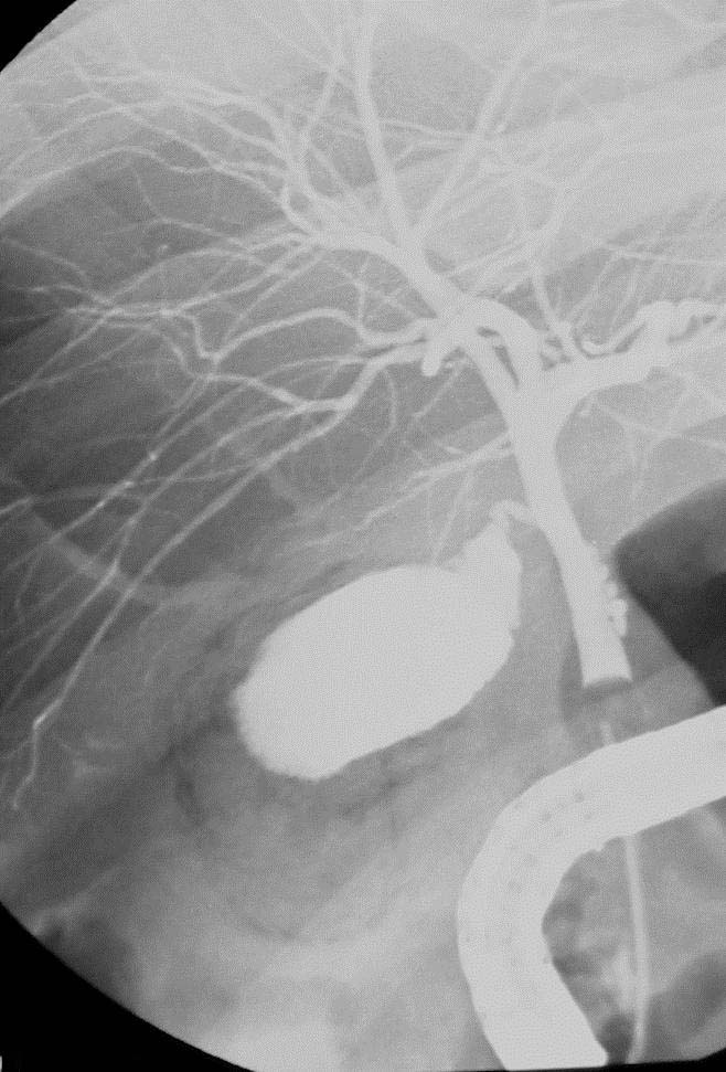
Cholangiography
Figures: Radiographic images of right upper quadrant of abdomen taken during ERCP examination. (A) Prior to filling of gallbladder lumen with contrast agent, gas (arrows) is noted lateral to the common bile duct.
(B) After filling of the gallbladder lumen, the gas is confirmed to be in the gallbladder wall.